Abdominal cysts can arise from a variety of underlying factors, ranging from congenital conditions to infections or trauma.
Their development often depends on the type of cyst and its location within the abdominal cavity.
Identifying the causes of these cysts is crucial in understanding their potential impact on health and the necessary steps for treatment.
Key Takeaways:
- Types of abdominal cysts are mesenteric cysts, intestinal duplication cysts, renal cysts, and splenic cysts.
- Ultrasounds and CT scans are pivotal for diagnosing all types of abdominal cysts, providing crucial details on size, location, and potential complications.
- Laparoscopic procedures are frequently used for many cyst types, offering benefits like quicker recovery and fewer complications.
- Potential issues include infections, organ dysfunction, or chronic pain, emphasizing the importance of early detection and management.
Mesenteric Cysts
Mesenteric cysts are fluid-filled sacs that form within the mesentery, the tissue that anchors the small intestine to the abdominal wall.
These cysts are more frequently observed in females than males and can range widely in size.
While some may remain asymptomatic and go unnoticed for years, others cause noticeable symptoms, particularly in children.
Common signs include abdominal pain, vomiting, and the presence of a palpable mass in the abdomen.
Key Characteristics of Mesenteric Cysts
- Location: Found in the mesentery of the small intestine.
- Prevalence: More common in males.
- Symptoms in children:
- Abdominal pain
- Vomiting
- Detectable mass in the abdomen
A notable concern with mesenteric cysts is their ability to cause volvulus, a condition where the bowel twists around itself. This leads to sharp abdominal pain, vomiting, and can rapidly escalate into a medical emergency.
The cysts can become infected, causing fever and a spike in white blood cell counts, further complicating the condition.
Diagnosis and Treatment
Now let us talk about diagnosis and treatment:
Diagnosis
Mesenteric cysts are typically identified through advanced imaging techniques, which provide critical insights into their size, location, and impact on nearby structures:
- Ultrasound: Provides detailed imaging of the cyst, offering precise measurements and determining its position within the mesentery.
- CT Scan: Offers a comprehensive view, assessing how the cyst interacts with surrounding organs and detecting any potential complications.
Treatment Options
- Surgical Removal:
- Frequently involves partial resection of the small bowel to eliminate the cyst.
- Minimally Invasive Laparoscopic Surgery:
- Promotes quicker recovery times.
- Reduces the likelihood of surgical complications.
Early diagnosis and the use of advanced surgical techniques play a crucial role in managing mesenteric cysts effectively, ensuring better long-term health outcomes.
Prompt identification and management of mesenteric cysts are essential to prevent severe complications, such as bowel obstruction or infection.
Early intervention not only mitigates immediate risks but also improves long-term outcomes for patients.
Intestinal Duplication Cysts

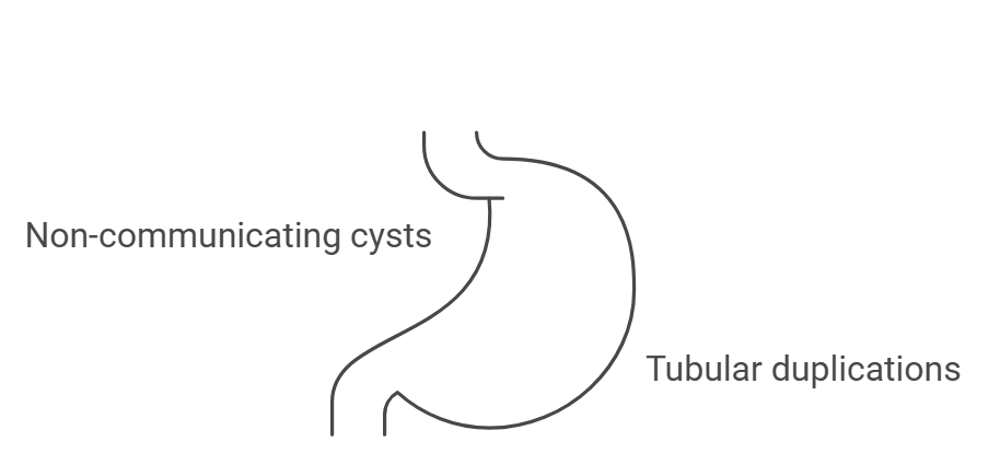
Intestinal duplication cysts, also referred to as enteric cysts, are rare congenital abnormalities that can form anywhere along the gastrointestinal tract, from the base of the tongue to the rectum.
The majority are found in the abdominal cavity, but some may develop in the thoracic region or even span across the diaphragm.
These cysts are classified into two main categories:
- Non-communicating cysts: Most duplication cysts do not connect to the bowel lumen and are isolated.
- Tubular duplications: Approximately 25% of cases involve tubular duplications that communicate directly with the bowel.
In some cases, the lining of these cysts contains ectopic tissues, such as gastric or pancreatic mucosa.
These tissues can produce digestive enzymes or acid, leading to complications such as:
- Ulceration
- Bleeding
Symptoms and Presentation
Symptoms of intestinal duplication cysts often manifest within the first two years of life.
- Partial intestinal obstruction, leading to abdominal pain or bloating
- Difficulty swallowing or breathing in cases where cysts are located in the thorax
- In rare cases, noticeable masses or other complications depending on the location
Diagnosis
Diagnostic imaging is crucial for identifying these cysts and planning treatment. Common imaging methods include:
- Ultrasound: Often used as the first-line diagnostic tool for its safety and clarity.
- CT Scans: Helpful for detailed visualization, particularly when multiple cysts are suspected.
- Since about 10% of patients may have more than one cyst, imaging of multiple body cavities (e.g., abdomen and thorax) is often recommended.
Treatment Options
Symptomatic cysts typically require surgical intervention. The goals of treatment are to relieve symptoms, prevent complications, and ensure a smooth recovery.
- Laparoscopic surgery: A minimally invasive option for abdominal cysts, reducing recovery time and surgical risks.
- Thoracoscopic surgery: Used for cysts located in the thorax, providing effective results with less postoperative discomfort.
Both methods allow for precise resection of the cyst while minimizing trauma to surrounding tissues. Post-surgery, patients generally recover quickly and with fewer complications compared to traditional open surgery.
Renal Cysts
Renal cysts can manifest in various forms, ranging from simple, benign structures to more complex and potentially life-altering conditions like multicystic dysplasia and polycystic kidney disease.
These cysts, depending on their type, have different risk factors, implications, and management strategies.
Simple Renal Cysts
Doctors often discover simple renal cysts, which are non-cancerous, fluid-filled sacs, incidentally during imaging studies performed for other reasons.
Although these cysts rarely cause symptoms, healthcare providers must carefully distinguish them from other conditions, such as:
- Hydronephrosis: A condition where urine buildup causes the kidney to swell.
- Cysts in neighboring organs: For example, in the liver or adrenal glands, which can sometimes resemble renal cysts.
Multicystic Dysplasia
Multicystic dysplasia is a congenital condition often identified prenatally through ultrasound. It is marked by the presence of multiple cysts of varying sizes within the kidney, with no identifiable functional kidney tissue. Key characteristics include:
- Abnormal kidney structure: The affected kidney is non-functional.
- Compensatory function: The unaffected kidney usually adapts and functions normally.
- Potential complications: Hypertension, urinary tract infections, or pain may arise, requiring surgical resection in severe cases.
This condition underscores the importance of prenatal imaging and postnatal monitoring to address any complications early.
Polycystic Kidney Disease (PKD)
Polycystic kidney disease is a hereditary condition involving the formation of numerous microscopic cysts in both kidneys.
These cysts can disrupt normal kidney function and lead to renal insufficiency, either at birth or later in life.
Associated Complications
- Pulmonary hypoplasia: Underdevelopment of the lungs, which can be life-threatening in newborns.
- Oligohydramnios: A deficiency of amniotic fluid that complicates pregnancy and affects fetal development.
- Progressive renal insufficiency: May lead to chronic kidney disease over time.
Management and Diagnosis
Timely diagnosis of renal cysts, especially during prenatal stages, is critical. Imaging techniques such as ultrasound and CT scans are invaluable in distinguishing between types of cysts.
Depending on the condition and its severity, management strategies include:
- Monitoring: For simple cysts that do not cause symptoms.
- Surgical intervention: For multicystic dysplasia or complicated cases involving infection or obstruction.
- Supportive care: For polycystic kidney disease, including measures to slow disease progression and manage symptoms like hypertension.
Splenic Cysts

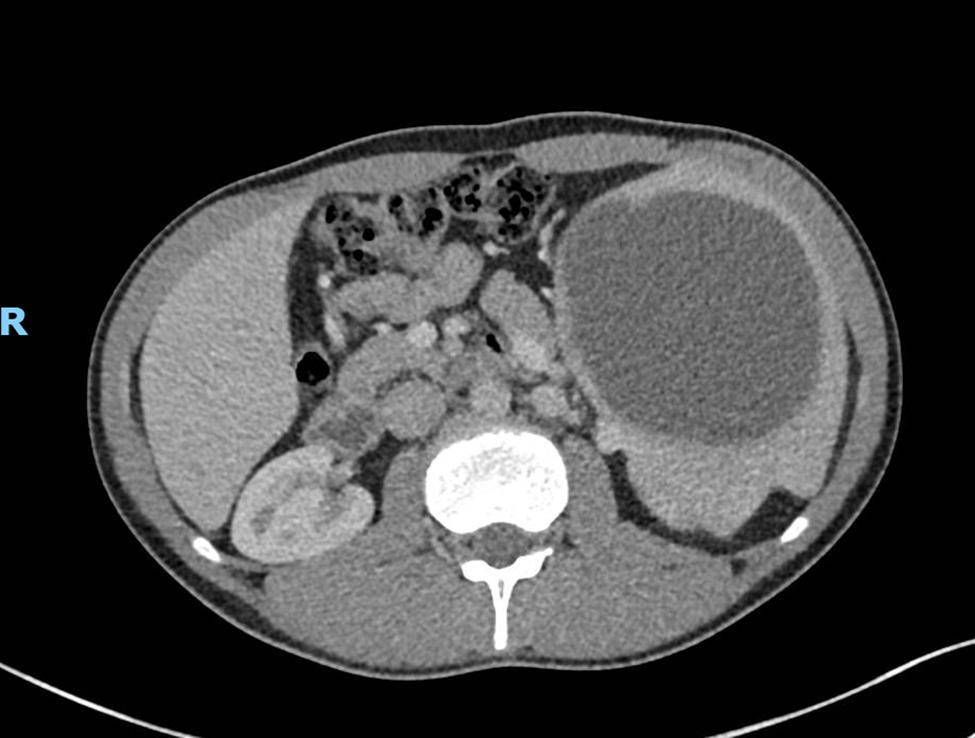
The next one we want to talk about is Splenic cysts, also known as pseudocysts, are abdominal cysts most often associated with previous trauma to the spleen.
The trauma can result from physical injuries, such as blunt force impact during accidents or falls, which cause damage to the spleen’s tissue and lead to cyst formation.
These cysts may remain asymptomatic for some time but often present with abdominal pain.
They are typically discovered when patients report a palpable mass in the left upper quadrant of the abdomen or during imaging studies conducted for other reasons.
- Abdominal pain.
- A noticeable mass in the left upper abdomen.
- Potential discomfort when sitting or lying in certain positions.
Cause: Frequently linked to previous injuries or trauma to the spleen.
Diagnosis is most reliably achieved through imaging techniques such as ultrasound or CT scans. These tools provide detailed images, allowing healthcare providers to differentiate splenic cysts from other potential issues like tumors or abscesses.
Treatment Options:
- Cyst Removal: Involves surgical excision of the cyst wall.
- Partial Splenectomy: For larger or more complex cysts, part of the spleen may need to be removed.
Minimally Invasive Surgery:
- Laparoscopic techniques are commonly used.
- Benefits include shorter recovery times, reduced scarring, and fewer surgical risks.
Potential Complications:
- Recurrent cyst formation, requiring ongoing medical monitoring.
- Rare cases of infection or rupture, which can lead to more severe health issues.
The Bottom Line
Abdominal cysts are caused by a variety of factors, including congenital anomalies, trauma, and infections.
Early identification through imaging and timely surgical intervention are key to preventing complications.
Awareness of these conditions ensures timely medical intervention and better long-term health outcomes.
